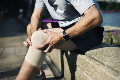
Dr. Eric Rush of SIMEDHealth's Rehabilitation Medicine recently wrote an article for the spring/summer edition of House Calls magazine, a seasonal publication by the Alachua County Medical Society. In this article, he discusses treatment plans for Osteoarthritis of the knee that do not involve surgery.
Check out the full article below:


Article content:
Background:
Arthritis is the leading cause of disability in the US with an estimated 52.5 million adults suffering from this condition (2). The CDC denotes arthritic joint pain that severely impacts a person’s quality of life as severe joint pain (SJP) and found that of the 52.5 million adults with documented arthritis over 27% of them suffered from SJP (2). Finding ways to help our patients to live with osteoarthritis (OA) and SJP is of paramount importance, especially in our home of Alachua county which has a median age of 31.3.
Diagnosis and Workup of Arthritic Knee Pain
The beginning of an arthritis diagnosis like any other medical condition starts with a thorough history and physical examination. Ruling out inflammatory conditions (gout, pseudogout etc.) and systemic spondyloarthropathies (RA, ankylosing spondylarthrosis etc.) is important when first making the diagnosis of primary knee OA. Red-Flag Symptoms that should make you think of other causes of knee pain include, fever, weight changes, many joints involved, erythema, swelling, warmth, prolonged morning stiffness (> 1 hour), and rapid onset of knee pain. Knees which are swollen, erythematous and red should always be considered for infection, and gouty arthropathies, and fluid analysis on the synovial fluid is critical in those diagnoses.
A classic OA picture will present as a gradual worsening of symptoms, initial stiffness in the am (usually < 30 minutes), worse after rest but improves somewhat with moderate exercise, can be provoked by prolonged activity. The patient may report clicking or popping but it is uncommon for the joint to lock or become stuck with just OA. The pain is usually aching and deep in nature but can have some occasional sharp pains reported. Typical OA will develop worse in the medial compartment of the knee with a slight valgus (knees buckled in) appearance. In younger patients (<55 years old) primary patellofemoral arthritis can cause severe pain which remains localized deep to the patella and is worse with extension type activities.
Before moving forward with treatment, it is important to perform a thorough physical exam. Inspection is often overlooked but is vitally important, deformities of the knee will cause uneven stresses and can lead to more rapid progression of the arthritis and more severe symptoms. Palpation along the joint lines of the knee as well as range of motion (ROM) of the knee can give insight into general function and localization of pain symptoms. Testing of the ligaments of the knees should include varus and valgus stress testing both in full extension and at 20-30* of flexion, as well as anterior and posterior stability testing with either an anterior and posterior drawer test or Lachman test. Gait observation including limp, and comparative stride length can give insight into the impact arthritis may be having on your patient.
Further testing with laboratory studies and imaging can be beneficial when first making the diagnosis of primary knee OA. Weight bearing XR of the knee in AP, Lat and NWB sunrise views gives the best images of the knee in its functional state and can better help to establish progression of disease. Routine laboratory studies with CBC, CMP, sed-rate and/or CRP may be warranted if the patient has any of the red flag symptoms above, or you are considering pharmacologic interventions for their pain, however lab testing is usually not necessary for diagnosis of OA..
Non-pharmacologic Management
The first steps for managing OA knee pain are lifestyle changes with an emphasis on diet and exercise. Moderate intensity exercise of at least 150 minutes per week has been shown in several studies to have beneficial effects on slowing the progression of and disability from knee OA (4). Also, BMI of higher than 35 carries a very sharp rise in the risk of infection post joint replacement, and thus it is important that from the first sign of joint pain a push is made to combat obesity (5). Physical Therapy with strengthening programs targeting the adductors and knee extensor muscles will help to stabilize the knee and assist in OA pain deriving from patellofemoral component arthritis. Regular stretching to preserve full ROM is also important as one of the most predictive factors of knee ROM post replacement is knee ROM pre-replacement. Complementary and alternative medicines such as acupuncture, heat and ice modalities, and herbal supplements have also been shown to have benefits in small studies.
Pharmacologic Management
Acetaminophen should be considered first line for oral pharmacologic treatment. Doses of 650mg up to four times daily can be effective in most early cases of OA. Prolonged use of acetaminophen should prompt the physician to check liver transaminases for possible adverse events with the medication. Oral NSAID or topical NSAIDs should be considered second line. Chronic use of either should prompt a physician to check renal function every 3-6 months as well as considering placing the patient on an antacid medication to reduce risk of gastrointestinal complications. Topical agents either via gel, patch or liquids carry less risk of the systemic side effects but have been less effective in overweight patients. Use caution in using oral NSAID or selective COX inhibitors in patients with known coronary artery disease as there is a significant increased risk of cardiovascular events with prolonged use.
Interventional Management
If pain continues despite pharmacologic and non-pharmacologic treatments, the next step in management is interventional modalities. There are several types of injections and a multitude of mechanisms of action that can lead to reduction in arthritic pain and morbidity. Arthrocentesis should be considered for every patient on their first injection to allow for fluid analysis to rule out concomitant gout or pseudogout which may play a role in worsening the patient’s prognosis. Depending on the severity of the arthritis and the level of effusion within the joint it may be beneficial to the patient to first perform an arthrocentesis, as the amount of fluid in the injection can lead to increased intraarticular pressure and discomfort.
After simple arthrocentesis the next most common injection includes intraarticular steroid injection with or without local anesthetic. I recommend using local anesthetic when injecting steroids as it can limit some of the discomfort from joint capsule stretch from injection of all the fluid. While more recent studies show that there may be some increase in the rate of cartilage degradation post corticosteroid injection, and that long term the injections do not delay or prevent joint replacement patients often report improved symptom and impairment with these injections (1,3). Alternative injections with hyaluronic acid or viscosupplementation has shown benefits in the management of OA, however it seems to be very patient specific and at this point no clear predictive indices for who will be a responder and who will not currently exist (3). For these injections improvement of symptoms >50% at 2 weeks and continued benefit past 6 months is considered a successful treatment, and you could consider repeat viscosupplementation injections. They come in various options for injection including single dose up to 5 sequential weekly injections.
The newer options for interventional management of knee OA pain include geniculate nerve blocks which operate on a principle like medial branch rhizotomies on the back. First the patient will have a diagnostic block or two to identify if the formal rhizotomy is likely to provide relief. Then if the patient reports >50% reduction in their pain symptoms which wears off appropriately for the anesthetic they will be brought back for a radio-frequency ablation of the geniculate nerves; thus, denervating the articular surfaces of the knee. No motor function is lost in this procedure as all motor branches have been given off prior to the site of the lesion. The remaining two options include platelet rich plasma (PRP), or stem cell injections in which the patient has cells harvested from autologous tissue and then injected into the knee. The mechanism of action of this is an inflammatory process and thus patients are instructed to stop all NSAIDs and steroids prior to injections. Both last two options are still classified under investigatory by most insurances and thus are usually performed as self-pay interventions.
Ultimately, the goal of the physician in treating chronic knee OA pain is to limit pain symptoms which impair the patient’s quality of life. Doing so in the least invasive way possible is the ultimate goal. There is no treatment currently that has been shown to prevent the progression of OA or reverse damage that has already been done, and our goal should be to delay replacement as far into life as possible to prevent the need of revision and to set patient’s up for success post replacement with strengthening, ROM, and weight loss strategies if they end up needing to undergo total knee replacement.
To request an appointment with Dr. Rush, click here